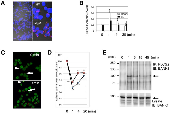
Figure 3. BANK1-PLCG2 complex formation is transient and induced by IgM stimulation.
(A) Confocal images of Daudi B cells showing increase molecular proximity between endogenous BANK1 and PLCg2 proteins upon anti-IgM stimulation. The staining was done using in situ a PLA protocol with rabbit anti-BANK1 (ET-52) and mouse anti PLCg2 (Abcam). Nuclei were stained with DAPI in blue. The confocal images (PLA signals) were taken with a pinhole of 2.5 (Zeiss Plan-Apochromat 63× oil objective). Upper panel, non-stimulated cells. Low panel, cells stimulated for 1 minute with anti-human IgM-F(ab')2. To the right are shown digital magnifications. (B) Time variation of PLA BANK1-PLCg2 interaction upon stimulation with anti-IgM in two human B-cell lines, the Burkitt´s derived Daudi and the non-Hodgkin´s lymphoma derived RL. Results are shown as the mean from three independent experiments. Error bars represent the SD from the mean. *P<0,05 based on Student´s test comparing stimulated cells versus time 0 (non stimulated cells).(C) Merge confocal images (pinhole = 1) of Daudi cells showing the nuclei in green and the BANK1-PLCg2 PLA interaction in red to permit co-location analysis. PLA signals close to the nucleus appeared as yellow dots (arrowheads) and when localize in the periphery of the cell as red dots (arrows). (D) Time course upon anti-IgM stimulation of co-localization between the PLA signal and the nucleus. Quantification was done using the overlap coefficient after Manders because this coefficient is insensitive to differences in signal intensities between channels. After one minute of stimulation the co-localization decreases suggesting a translocation of the BANK1-PLCg2 complex. The graft shows two independent experiments, each point represents the analysis of at least 300 cells.**,P<0,01;***P<0,001 based on Student´s test comparing stimulated cells versus time 0 (non stimulated). (E) Immunoprecipitation of the BANK1-PLCg2 complex in Daudi cells. Anti-PLCg2 immunoprecipitates (above) and total cell lysate (below) were analyzed by immunoblotting with anti-BANK1 antibody (BANK1-ET-52). The position of the BANK1 protein is indicated by arrows.

