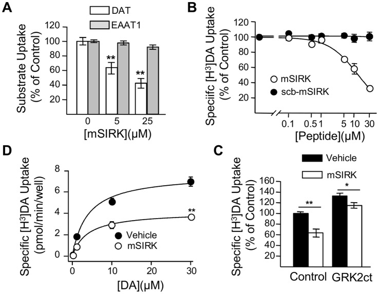
Figure 5. Activation of Gβγ with mSIRK resulted in an inhibition of DAT activity in heterologous systems.
(A) Oocytes expressing DAT (white bars) or EAAT1 (gray bars) were incubated for 30 min with 0.5% DMSO (DAT/n = 3; EAAT1/n = 25), 5 µM mSIRK (DAT/n = 20, EAAT1/n = 23), or 25 µM mSIRK (DAT/n = 15, EAAT1/n = 15) before uptake assays with [H3]-DA or [3H]-Glutamate, respectively. (B) Dose-dependent inhibition of [3H]-DA uptake in HEK293-DAT cells by mSIRK (n = 4, white circle). (C) 5 min pre-incubation of HEK293-DAT cells with 10 µM mSIRK produced a reduction of Vmax with no changes in Km (n = 3). (D) In HEK293-DAT, 10 µM mSIRK reduced uptake (control, black bar, n = 32; mSIRK, white bar, n = 15). The inhibitory effect of 10 µM mSIRK was attenuated in HEK-DAT cells expressing GRK2ct (control, n = 16; mSIRK, n = 16). **p<0.01 or *p<0.05.

