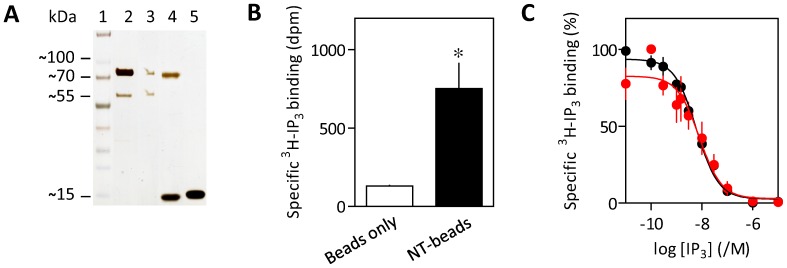
Figure 3. Immobilization of functional NT on beads.
(A) SVA-beads (60 pmol) were incubated with NT-biotin (17 pmol) and then magnetically separated from the supernatant (see Methods). The silver-stained gel shows equivalent fractions of the input (lane 2), the supernatant (lane 3), NT-beads treated (85°C, 10 min) to release bound protein (lane 4), or similarly treated control beads (lane 5). Lane 1 shows the molecular mass markers (kDa). The 70-kDa- and 15-kDa-bands correspond to NT-biotin and SVA monomer, respectively. (B) Specific 3H-IP3 binding (0.75 nM) to control or NT-coated SVA-beads. Results (dpm, disintegrations per minute) are means ± SEM, n = 3. *P = 0.019. (C) Equilibrium-competition binding to NT-biotin (black) and NT-beads (red) with 3H-IP3 (0.75 nM). Results are means ± SEM from 3 experiments.

