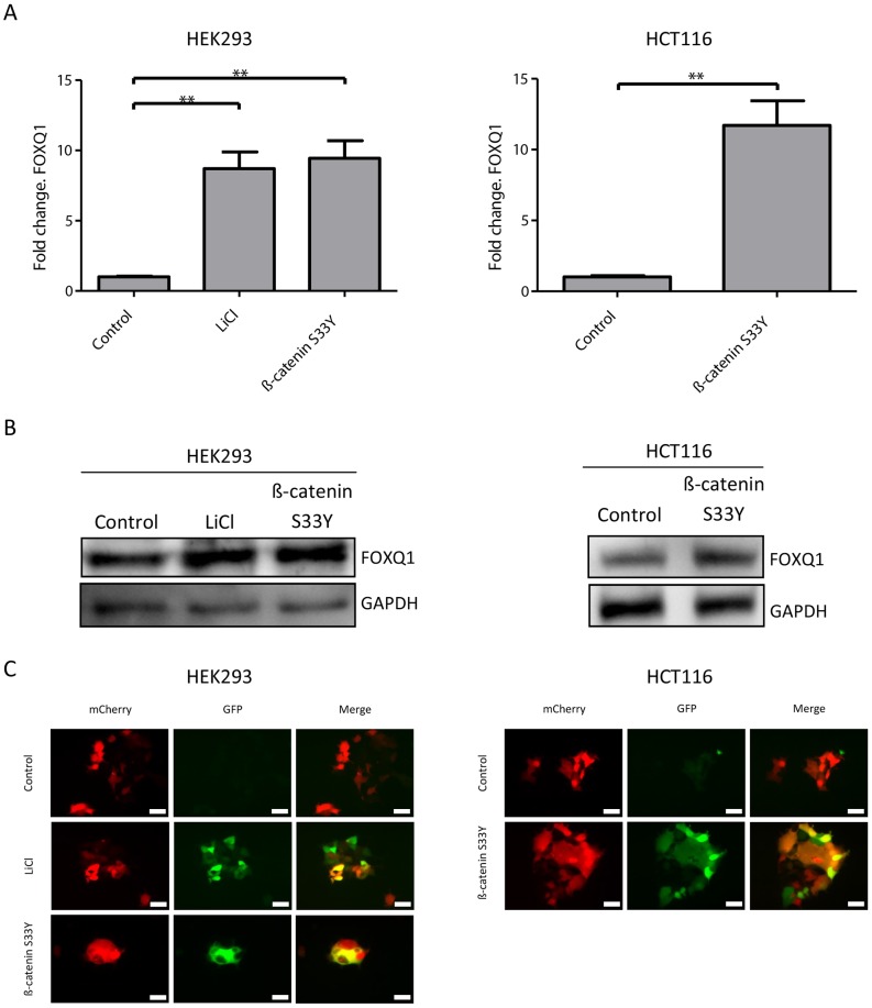
Figure 5. Activation of the Wnt pathway leads to increased FOXQ1 expression.
(A) qRT-PCR analysis of FOXQ1 expression in HEK293 and HCT116 cell lines after Wnt activation. Cell were treated with 20 mM LiCl for 24 hours or transfected with a constitutively active form of ß-catenin (S33Y). Data are mean ± SD n = 3, **P<0.01 using Mann-Whitney test. (B) Western blot of FOXQ1 in HEK293 and HCT116 after Wnt activation as described above. GAPDH was used a loading control. (C) HEK293 and HCT116 cells stably expressing the Wnt sensitive reporter 7TGC [25] were treated as described above and imaged. GFP (green) is under the control of a Wnt sensitive promoter and mCherry (red) is constitutively expressed to identify infected cells, white bar indicates 20 µm.

