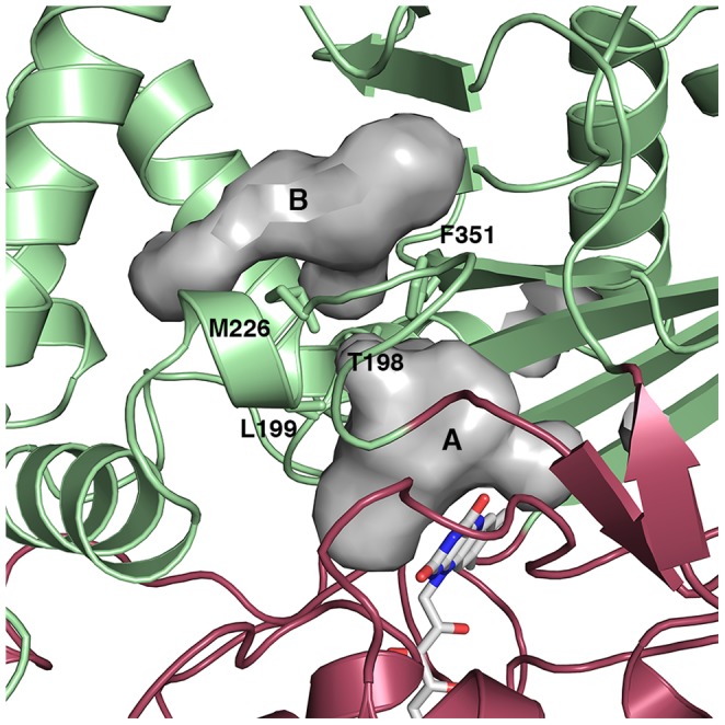
Figure 2. Access to the CHAO active site.

An illustration of the substrate-binding (A) and secondary (B) cavities in the CHAO structure. The protein is shown as a cartoon figure, with the substrate-binding domain colored green and the cofactor-binding domain colored red. FAD is shown as sticks, as are four residues that separate the two cavities (T198, L199, M226, and F351). Cavities were visualized with the program PyMOL using the default 1.4 Å probe radius.
