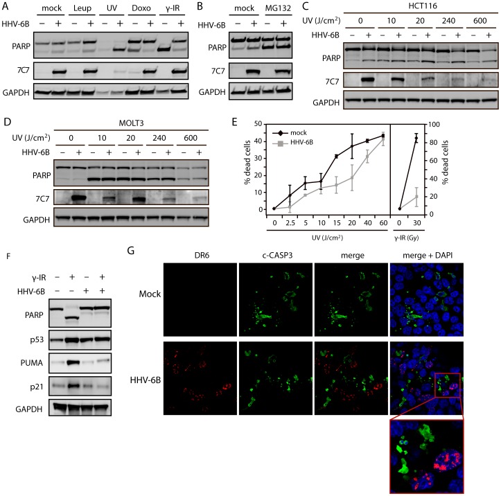
Figure 2. HHV-6B infection rescues cells from p53-dependent apoptosis.
(A & B) Western blot analysis of PARP cleavage in cells either mock-treated or infected with HHV-6B for 48 hrs. The cells were subsequently treated with mock, leupeptin (10 µM), UV radiation (10 min exposure followed by 4 hrs of incubation), doxorubicin (0.2 µg/ml for 24 hrs), γ radiation (30 Gy followed by 24 hrs of incubation), or MG132 (10 µM). 7C7 was used as infection control and GAPDH was used as loading control. (C & D) Western blot analyses with antibodies against PARP, GAPDH, and 7C7 (infection control) on lysates from HCT116 (C) or MOLT3 (D) cells treated with varying doses of UV radiation (10, 20, 240, 600 J/cm2 followed by 4 hrs of incubation). (E) ATP cell viability assay on MOLT3 cells with or without HHV-6B infection and treated with varying amounts of UV (0, 2.5, 5, 10, 15, 20, 40 and 60 J/cm2 followed by 4 hrs of incubation) or γ radiation (0 and 30 Gy followed by 24 hrs of incubation). (F) Western blot analyses with antibodies against PARP, p53, PUMA, p21 or GAPDH (loading control) on lysates from HCT116 cells either mock-treated or infected with HHV-6B for 24 hrs followed by γ radiation (30 Gy) and 24 hrs of incubation. (G) Confocal microscopy images of HCT116 cells either mock-treated or infected with HHV-6B for 24 hrs, γ-irradiated (30 Gy) followed by 24 hrs of incubation. The cells were analyzed for cleaved caspase-3 (Alexa 488 green), DR6 (Alexa 546 red), and DAPI (blue).

