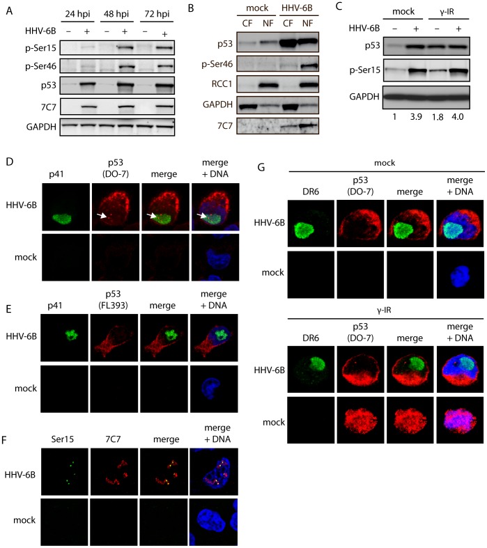
Figure 3. HHV-6B infection leads to nuclear accumulation of phosphorylated p53.
(A) Western blot analyses of HCT116 cells infected for 24, 48, or 72 hrs and analyzed with antibodies against p53 p-Ser15, p53 p-Ser46, p53, 7C7 (infection control), or GAPDH (loading control). (B) Western blot analyses of nuclear (NF) and cytoplasmic (CF) fractions from mock-treated or HHV-6B-infected HCT116 cells (48 hpi). The membranes were stained with antibodies against p53, p53 p-Ser46, 7C7 (infection control), RCC1 (nuclear control), or GAPDH (cytoplasmic control). (C) Western blot analyses of HCT116 cells mock-treated or HHV-6B-infected for 24 hrs followed by γ irradiation (30 Gy) and additional incubation for 24 hrs. The membrane was probed with antibodies against p53, p53 p-Ser15, or GAPDH (loading control). Numbers at the bottom of the figure indicate fold induction of Ser15 phosphorylation relative to GAPDH. (D & E) Confocal microscopy on MOLT3 cells infected with HHV-6B for 24 hrs followed by staining with antibodies against the viral protein p41 (green), p53 (red) DAPI (blue). The p53 antibodies were either the monoclonal DO-7 (D) or a polyclonal antibody (FL393) (E). Arrows point to nuclear p53 staining. (F) Confocal microscopy on MOLT3 cells infected with HHV-6B for 24 hrs followed by staining with antibodies against p53 phospho-Ser20 (green), 7C7 (red) and DAPI (blue). (G) Confocal microscopy on MOLT3 cells infected with HHV-6B for 24 hrs, γ-irradiated (30 Gy) for 24 hrs followed by staining with antibodies against the viral protein DR6 (green), p53 (DO-7) (red) DAPI (blue).

