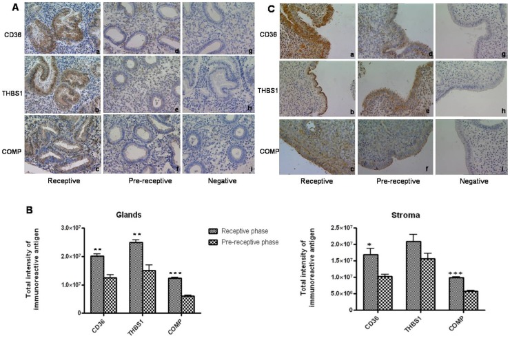
Figure 5. Immunolocalization of the proteins encoded by select RAGs in receptive and pre-receptive phase human endometrial tissues.
A) Immunohistochemical localization of CD36, THBS1 and COMP in receptive phase (a,b,c) and pre-receptive phase (d,e,f) endometrial tissues. Panels g and i represent the sections stained with rabbit IgG and h shows the section stained with mouse IgG. B) Semiquantitative analysis to compare the intensities of immunoreactive CD36, THBS1 and COMP in epithelial and stromal compartments of receptive and pre-receptive phase human endometrial samples. *p<0.05, ** p <0.005, *** p<0.002 C) Immunohistochemical localization of CD36,THBS1 and COMP in the luminal epithelium of human endometrial tissues collected in receptive phase (a,b,c) and pre-receptive phase (d,e,f) of menstrual cycle. Panels g and i represent the sections stained with rabbit IgG and panel h is stained with mouse IgG. Magnification at 40X.

