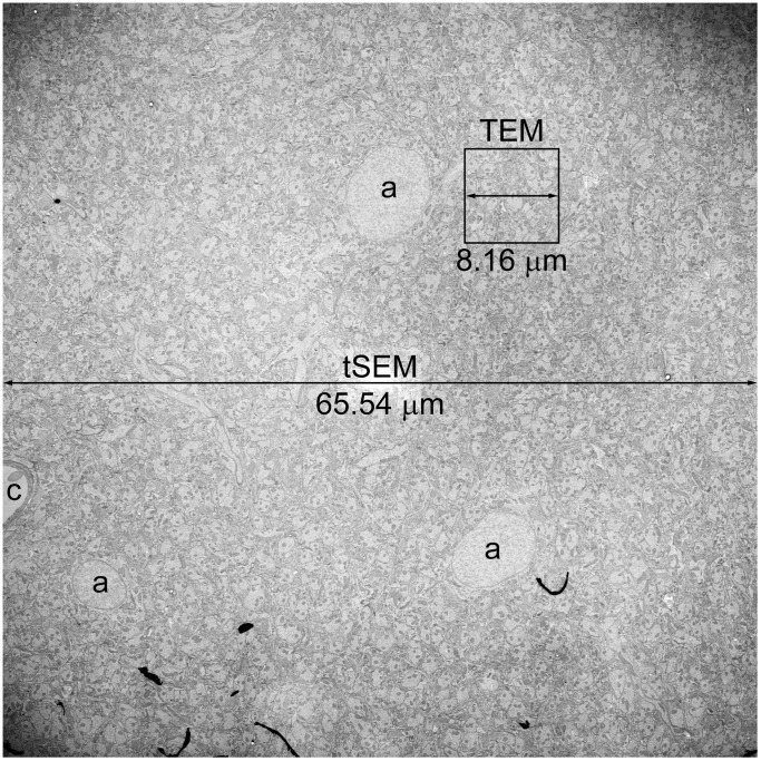
Figure 5. Field size comparison of images acquired on tSEM vs. TEM.
This single field image of the rat hippocampal dentate gyrus (inner molecular layer) was acquired on tSEM originally at 32,768×32,768 pixels, or 65.54 µm×65.54 µm at 2 nm/pixel. Three astrocyte soma with round nuclei (a) and part of a capillary (c) are visible in this image. Boxed area indicates the size of a single field that can be imaged on our TEM (4,080×4,080 pixels, or 8.16 µm×8.16 µm at 2 nm/pixel). Note that the size of TEM field is similar to that of the nucleus of an astrocyte. The image has been adjusted for brightness and contrast, and re-sampled from the original pixel dimensions to 1,836×1,836 pixels during preparation of this figure.

