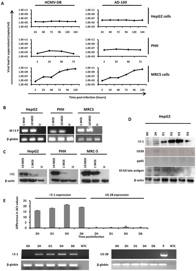
Figure 1. Growth curves of HCMV in HepG2 cells and PHH.
(A) Growth curves of HCMV in HepG2 cells, PHH and MRC5 cells. HepG2 cells, PHH, and MRC5 cells were left uninfected or infected with HCMV strains AD169 and HCMV-DB (MOI = 1). Inocula were left in place for 2 hours and then removed with three washes of EMEM without serum. Viral titers were determined in the culture supernatants at the indicated times post-infection by real-time PCR. Results are representative of two independent experiments. (B) Viral entry into HepG2 cells, PHH and MRC5 cells. Uninfected cells and cultures infected with HCMV-AD169 at the indicated MOI for 2 hours were treated with trypsin for 10 min and then washed. Samples of extracted DNA were analyzed by PCR using primers specific for the MIEP of HCMV and for beta-globin (internal loading control). The amplification products were resolved by 2% agarose gel electrophoresis and visualized by ethidium bromide staining. Results are representative of two independent experiments. (C) Detection of IE1 pp72 HCMV antigen in infected HepG2 cells and PHH. HepG2 cells (6×10⁶ cells) and PHH (2×10⁶ cells) were left uninfected or infected with HCMV-AD169 (MOI = 1 and 10). IE1 pp72 HCMV antigen expression was measured at day 3 post-infection by Western blotting as described in the Materials and Methods section. beta-actin was used as control. Results are representative of two independent experiments. (D) Detection of IE1 pp72, but not US28, pp65 antigen and 65-kD structural late antigen in infected HepG2 cells. HepG2 cells (6×10⁶ cells) were left uninfected or infected with HCMV-AD169 (MOI = 1). IE1 pp72, US28, pp65 and 65-kD structural late HCMV antigen expression was measured up to day 4 post-infection by Western blotting as described in the Materials and Methods section. beta-actin was used as control. Results are representative of two independent experiments. (E) Detection of IE1 pp72 transcript, but not of US28 transcript, in HCMV-infected HepG2 cells. HepG2 cells (6×10⁶ cells) were left uninfected or infected with HCMV-AD169 (MOI = 1). IE1 pp72 and US28 transcript expression was measured up to day 6 post-infection by RT-PCR assay as described in the Materials and Methods section. beta-globin was used as control. Results represent means (± SD) of two independent experiments. IE: Immediate Early; MIEP: Major immediate-early promoter; MOI: Multiplicity of infection; PHH: Primary human hepatocytes; U: Uninfected; P: positive control (extract of MRC5 cells infected with HCMV-AD169).

