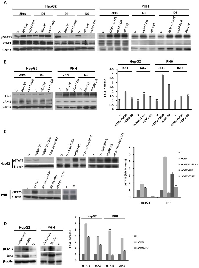
Figure 3. HCMV induces IL-6-mediated activation of the JAK-STAT3 axis in HepG2 cells and PHH.
(A) Time course of STAT3 activation in HepG2 cells and PHH infected with HCMV. HepG2 cells (6×10⁶ cells) were left uninfected or infected with HCMV strains AD169 and HCMV-DB (MOI = 0.5). PHH (2×10⁶ cells) were left uninfected or infected with HCMV strains AD169 and HCMV-DB (MOI = 1). STAT3 activation was measured by Western blotting as described in the Materials and Methods section. Unphosphorylated STAT3 and beta-actin were used as controls, and ganciclovir was used at a concentration of 5 microg/ml. (B) Time course of JAK1/JAK2 activation in HepG2 cells and PHH infected with HCMV. HepG2 cells (6×10⁶ cells) were left uninfected or infected with HCMV strains AD169 and HCMV-DB (MOI = 0.5). PHH (2×10⁶ cells) were left uninfected or infected with HCMV strains AD169 and HCMV-DB (MOI = 1). JAK1/JAK2 activation was measured by Western blotting, and beta-actin was used as an internal control. The histogram shows JAK activation at 2 hours post-infection as quantified using Image J 1.40 software. (C) STAT3 activation is mediated by the IL-6-JAK pathway in HepG2 cells and PHH infected with HCMV. HepG2 cells (6×10⁶ cells) and PHH (2×10⁶ cells) were left uninfected or infected with HCMV (MOI = 0.5) in the presence or absence of a JAK inhibitor (1 micromol/l), a STAT3 inhibitor (10 micromol/l), a neutralizing anti-IL-6R mAb (10 microg/ml), and a neutralizing anti-EGFR mAb (20 microg/ml). Cells were left uninfected or incubated with the recombinant HCMV glycoprotein gB (10 microg/ml) for 2 hours. STAT3 activation was measured by Western blotting at day 1 post-infection in PHH incubated with JAK inhibitor, STAT3 inhibitor, anti-IL-6R mAb, and in HepG2 cells incubated with JAK and STAT3 inhibitors. STAT3 activation was measured at 2 hours post-infection in HepG2 cells incubated with anti-IL-6R mAb and anti-EGFR mAb. beta-actin was used as an internal control. The histogram shows STAT3 activation as quantified using Image J 1.40 software. (D) STAT3 activation is mediated primarily by HCMV in HepG2 cells and PHH. HepG2 cells (6×10⁶ cells) and PHH (2×10⁶ cells) were left uninfected or infected with HCMV or UV-inactivated HCMV (AD169, MOI = 1). The activation of STAT3 and JAK2 was measured by western blot at day 3 post-infection. beta-actin was used as a control for equal loading. The histogram shows STAT3 and JAK2 activation as quantified using Image J 1.40 software. Results of western-blots are representative of two independent experiments; histograms represent means (± SD) of two independent experiments. Ab: Antibody; EGFR: Epidermal growth factor receptor; GCV: ganciclovir.

