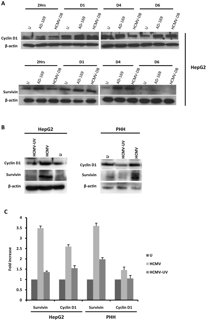
Figure 4. Up-regulation of cyclin D1 and survivin in HepG2 cells and PHH infected with HCMV.
(A) Time course of the expression of cyclin-D1 and survivin in HepG2 cells infected with HCMV. HepG2 cells (6×10⁶ cells) were left uninfected or infected with HCMV strains AD169 (MOI = 0.5) and HCMV-DB (MOI = 1.0). Cyclin D1 and survivin expression was measured by Western blotting as described in the Materials and Methods, and beta-actin was used as an internal control. (B) Expression of cyclin-D1 and survivin in PHH and HepG2 cells infected with live HCMV or UV-inactivated HCMV. HepG2 cells (6×10⁶ cells) and PHH (2×10⁶ cells) were left uninfected or infected with HCMV or UV-inactivated HCMV (AD169, MOI = 0.5). Cyclin D1 and survivin expression was measured by Western blotting as described in the Materials and Methods, and beta-actin was used as an internal control. (C) Expression of cyclin D1 and survivin is mediated primarily by HCMV in HepG2 cells and PHH. The histogram shows survivin and cyclin D1 expression at day 3 post-infection as quantified using Image J 1.40 software. Results of western-blots are representative of two independent experiments; histogram represents means (± SD) of two independent experiments.

