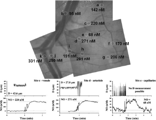
FIGURE 5.
Mapping of perivascular nitric oxide (NO) in superfused rat mesentery and small intestine conducted in our laboratory (unpublished) using Nafion-coated recessed NO microelectrodes (top panel). Examples of experimental vessel diameter measurements and NO measurements also are shown (bottom graphs). For venule and arteriole measurements, the NO microelectrode initially was positioned far from the vessel (zero reading), then moved to touch the outer surface of the vessel gently (perivascular NO value). The microelectrode image interfered with video diameter measurements when it was near the vessel (for clarity, the diameter signal is removed just before the tip touches the vessel). For the capillary-perfused site, the NO microelectrode was initially far from the tissue surface, then was moved to the surface and back out into the superfusion bath. The NO microelectrode current was converted to concentration based on calibrations at known NO concentrations.

