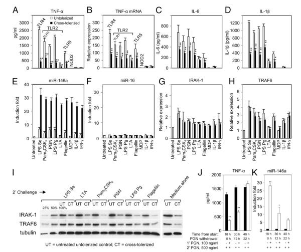
FIGURE 3.
High levels of LPS-induced miR-146a may account for cross-tolerance to non-LPS agonists in the THP-1 cell model. THP-1 cells primed with 10 ng/ml LPS for 18 h (cross-tolerized, filled bars) and untreated controls incubated for the same time period (untolerized, open bars) were washed twice with PBS, then challenged with various ligands or medium alone for 5 h. Culture supernatants and total RNA were analyzed for cytokines using ELISA (A, C, D), or miRNA (E, F, J) and mRNA expression (B, G, H) by qRT-PCR, as described in Materials and Methods. Changes in IRAK-1 and TRAF6 protein levels in LPS-primed and unprimed THP-1 cells challenged with various TLR ligands or medium alone for 2 h were analyzed by Western blot with tubulin expression shown as loading controls (I). Serial dilutions of untreated THP-1 cell lysates (100, 50, 25%) were included in the Western blot to document the semiquantitative measurement for IRAK-1 and TRAF6 expression. THP-1 monocytes were cultured with or without 100 ng/ml PGN continuously for 18 h followed by washing and cultured in complete medium for another 0, 12, or 22 h (PGN withdrawal). At each time point, 6 × 10⁵ cells were challenged with 500 ng/ml PGN for 3 h prior to analysis of TNF-α production by ELISA (J) and miR-146a expression by qRT-PCR analysis (K). All results are expressed as mean ± SD from three independent experiments. *p < 0.05; **p < 0.01 compared with untolerized THP-1 cells.

