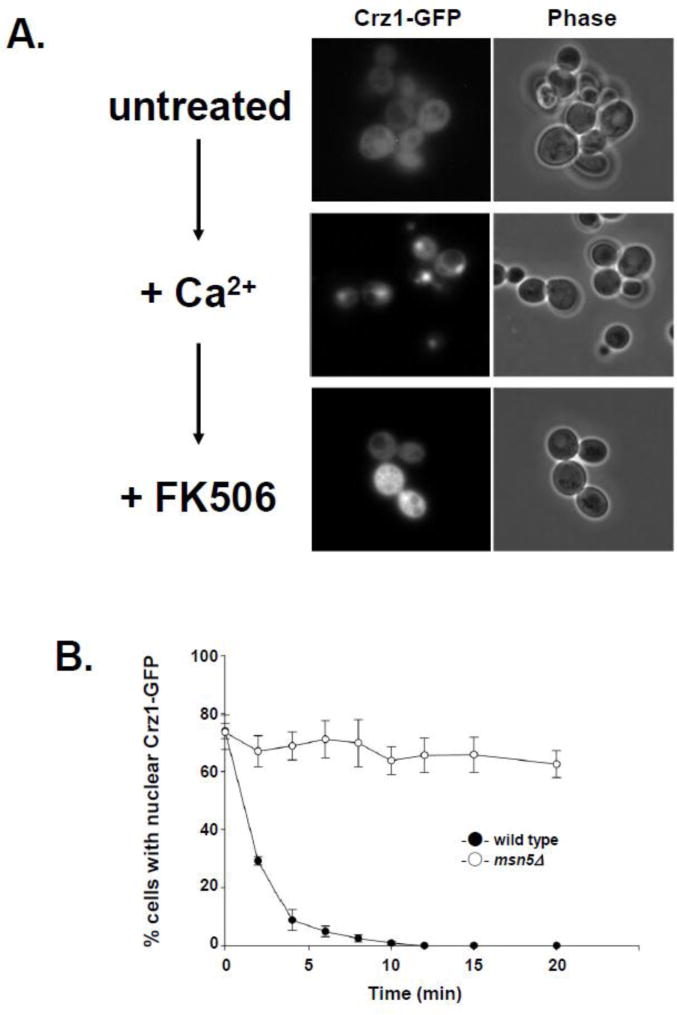
Figure 1.
Msn5-mediated nuclear export kinetics can be followed in vivo using FK506 induced nuclear export of Crz1-GFP from the nucleus. (A) Crz1-GFP localization changes upon addition of Ca2+ and FK506. Yeast expressing Crz1-GFP were examined using direct fluorescence microscopy prior to calcium addition (untreated), after the addition of CaCl2 to 170 mM for 5 min (+Ca2+), and after addition of FK506 to 1.4 ug/ml for 10 min (+FK506). Fluorescence (Crz1-GFP) and phase-contrast (phase) photomicrographs are shown. (B) Wild type and msn5Δ cells export Crz1-GFP at different rates following addition of FK506. Data points represent the mean percentage of cells with nuclear Crz1-GFP fluorescence at each time point based on a minimum of three assays. Error bars represent standard error of the mean.

