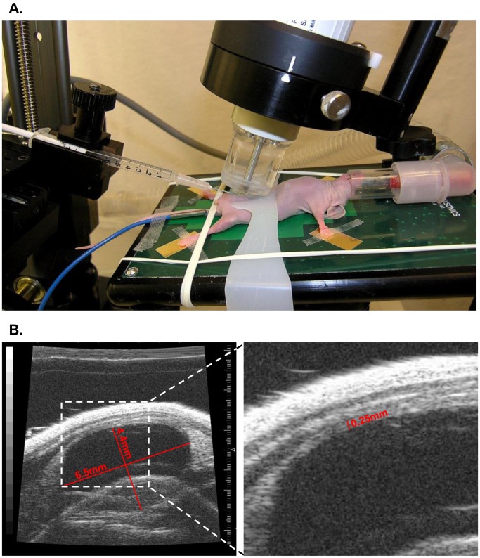
Figure 1. Ultrasound imaging.
A. The mice were anesthetized with isoflurane and mounted on the heated imaging table with continuous monitoring of vital signs. After visualization of the bladder with the Vevo 700® small animal imaging platform the skin was perforated with a 30G needle. B. Ultrasound visualisation of normal mouse bladder in sagittal section with typical dimensions indicated (lumen dimensions 4.4×6.5 mm; wall thickness 0.25 mm).

