
Figure 4. Neutrophils are recruited to the lungs in response to Lactobacillus priming.
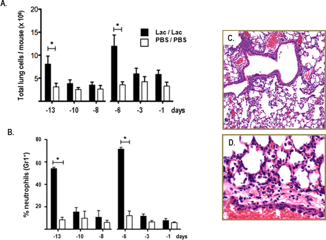
Cells from whole lung tissue evaluated by flow cytometry at three time points after the first (at day -14) and second (at day -7) inoculations with L. reuteri (Lac; filled bars) or PBS/BSA (PBS; open bars); A. Total lung cells B. Percent Gr1+ (neutrophils); n = 3 mice pooled for each time point, data compiled from 3 – 5 independent experiments; *p < 0.05; C. & D. Formalin-fixed, hematoxylin and eosin (H&E)-stained lung tissue from day -6, after second L. reuteri inoculation (see Fig. 1A); original magnifications, 10X and 40X, respectively.