FIGURE 6.
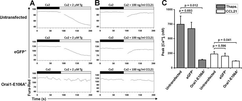
Inhibition of Ca2+ signals evoked by Tg or CCL21 treatment in human T cells expressing Orai1-E106A. A, Flow cytometry analysis, using the FlowJo Kinetics tool, of human T cells loaded with fura red. After 90 sec to establish a baseline, cells were treated with 2 μm Tg. Mean fluorescence changes (lower fluorescence = higher (Ca2+)i) are shown in untransfected human T cells (top), eGFP+ human T cells (middle), and eGFP-Orai1-E106A+ human T cells (bottom). B, Fluorescence changes in untransfected human T cells (top), eGFP+ human T cells (middle), and eGFP-Orai1-E106A+ human T cells (bottom) following addition of 100 ng/ml recombinant mouse CCL21. Data are from one donor and are representative of three donors. C, Mean peak [Ca2+]i signals evoked by Tg and CCL21 in control and eGFP-Orai1-E106A+ human T cells.

