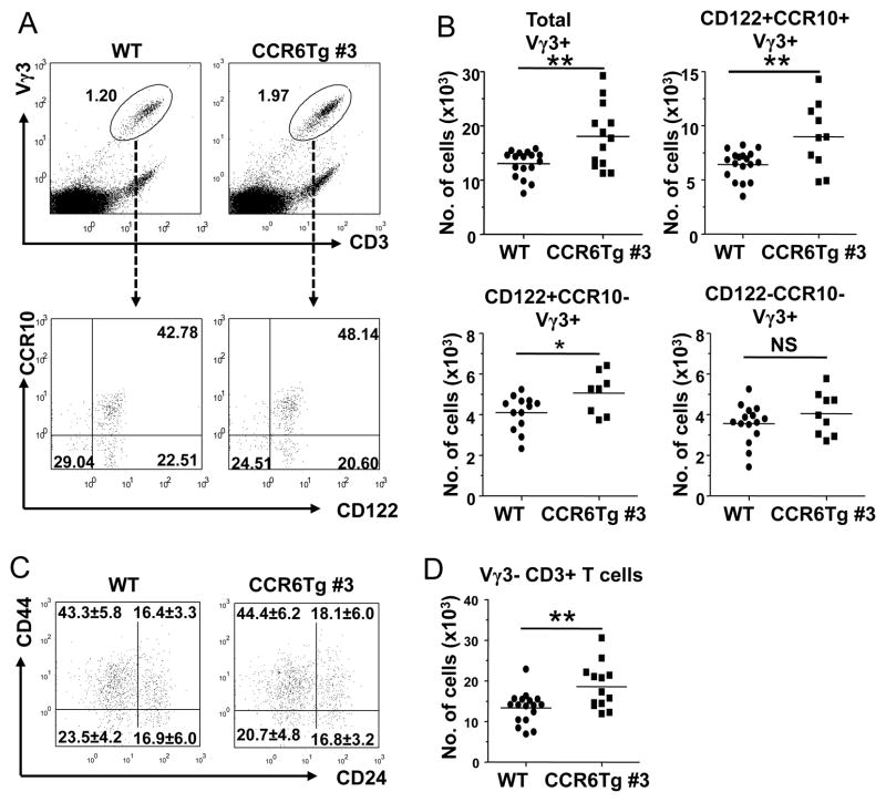
Figure 4.
Abnormal accumulation of mature Vγ3+ sIEL precursors in the fetal thymus of CCR6 transgenic mice. A. Flow cytometric analysis of E17 fetal thymocytes for Vγ3+ T cells of CCR6 transgenic and wild type littermates and their expression of CD122 and CCR10 (EGFP). B. Quantitative comparison of numbers of Vγ3+γδT cells of different maturation stages (total, CD122+CCR10+, CD122+CCR10− and CD122−CCR10−) in E17 fetal thymi of CCR6 transgenic and wild type littermates. Numbers of the different Vγ3+γδT cell subsets were calculated from total numbers of fetal thymocytes and the percentages of Vγ3+ fetal thymic γδT cells of different developmental stages as determined in the panel A. One dot represents the number of cells from one mouse. C. Flow cytometric analysis of E17 fetal thymic Vγ3+γδT cells of WT and CCR6Tg littermates for expression of CD44 and CD24. The cells in the histogram are of the gated Vγ3+CD3+ population. The numbers in each quadrant are average percentage ±standard deviation of cells in the quadrant. At least 9 mice of each genotype were analyzed. D. Quantitative comparison of numbers of E17 fetal thymic Vγ3− T cells in CCR6 transgenic and wild type littermates. The numbers were calculated similarly as in the panel B.

