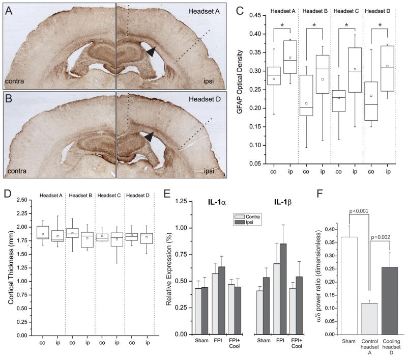
Figure 3. Prolonged mild focal cooling does not induce pathophysiological changes.
GFAP-immunostained coronal sections obtained from injured rats treated with control headset A (A) or cooling headset D (B). In A–B, arrows indicate enhanced GFAP+ staining characteristic of the white-matter/layer VI region ipsilateral to the injury; dotted lines delimit the approximate ipsilateral regions of interest. C) Box plots summarize the blind densitometric analysis of GFAP immunostaining. Immunoreactivity was significantly greater ipsilateral to the injury in each treatment group but no treatment differences were detected. All asterisks indicate p<0.05. Hollow square=mean; Whiskers indicate max and min values. Contra=contralateral, ipsi=ipsilateral to the injury site. D) Box plots summarize the blind analysis of neocortical thickness showing no significant effects of cooling. No differences were observed either between ipsilateral and contralateral neocortices within each group, or between ipsilateral neocortices across groups. In C–D co=contralateral, ip=ipsilateral. E) Three weeks of treatment with cooling headset D do not increase inflammation of the perilesional and homologous contralateral neocortices, as demonstrated by nominally lower gene expression levels of inflammatory cytokines IL-1α and IL-1β compared to headset A controls and sham-injured animals. F) Three weeks of cooling restore the perilesional ECoG power spectrum of stage N2 sleep. Compared to sham injured animals, a reduction of the alpha/delta ratio is evident in mock-cooled (headset A) rpFPI animals. This is significantly increased in cooled (headset D) rpFPI animals relative to randomized headset A.

