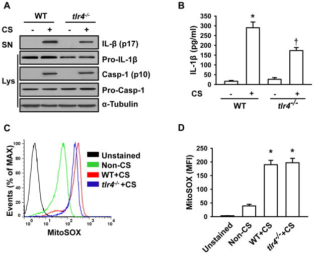
FIGURE 7. IL-1β production following cyclic stretch occurs via TLR4 signaling. A.
AMs isolated from tlr4−/− mice show less IL-1β release following stretch. Wild type and tlr4−/− AMs were subjected to 20% cyclic stretch for 4 h. IL-1β release in the culture supernatants (SN) of AMs and levels of Pro-IL-1β, Pro-Casp-1 and Casp-1 in cell lysate (Lys) were determined by Western blot analysis. B. The levels of IL-1β in cell-culture media were measured by ELISA (n=3). C. Representative histograms of flow cytometry experiments demonstrating the effects of cyclic stretch on mitochondrial ROS generation in tlr4−/− and wild type AMs. D. Quantitative data showing changes in mean fluorescent intensity (MFI) of MitoSOX following stretch (n=3). *p < 0.05 vs. the control (static) group. † p <0.05 vs. WT+CS group.

