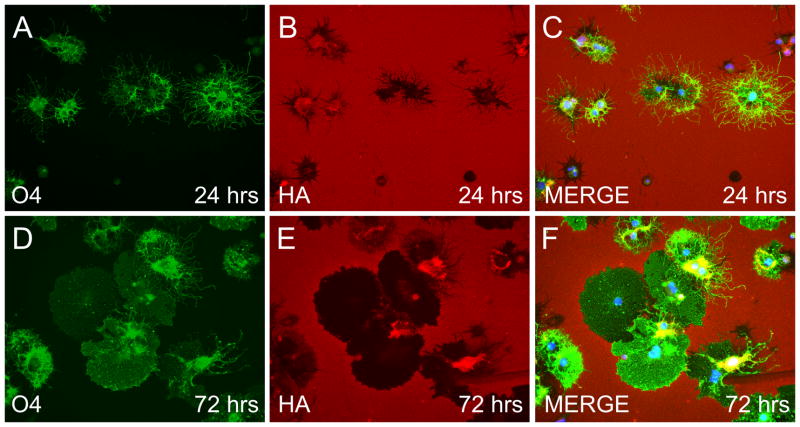
Figure 6.
OPCs and later OL stage cells degrade HA. OPCs grown in the presence of T3 and NAC were plated onto coverslips coated with HMW HA, allowed to mature for 24 (A–C) or 72 (D–F) hours and stained with O4 (green), HABP (red) and DAPI (blue). Pronounced degradation of HA, as assayed by loss of HABP staining, was seen around cell bodies and the extensive O4+ processes (A–C) at 24 hours. By 72 hours substantial degradation of HA was also observed (D–F) in areas corresponding to the presence of O4+ membranes that resemble myelin sheets. Note that HA was preserved around many OPC and mature OL cell bodies.

