Figure 8.
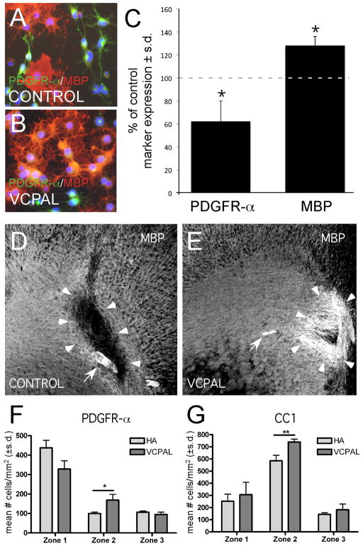
Inhibiting endogenous hyaluronidase activity promotes OL maturation in vitro and remyelination in vivo. (A) OPCs were grown in OL differentiation medium alone or (B) with 25μM VCPAL for 72–96 hours and stained with MBP, PDGFR-α and DAPI. Total MBP+ and PDGFR-α+ cells are quantified in (C). Consistent with the hypothesis that hyaluronidases expressed by OPCs generate HA digestion products that inhibit OL maturation, blocking HYAL activity with VCPAL increased the total percentage of MBP+ cells compared to controls (69% vs. 54% p= 0.005) while decreasing the total percentage of PDGFR-α+ cells (19% v. 30% in control, p=0.02). (D, E) MBP immunoreactivity (white) of lysolecithin lesions treated with HMW HA and vehicle (D) or HMW HA with 25 μM VCPAL (E). Arrowheads indicate the borders of the demyelinated lesion in D and the area of remyelination in E; arrows indicate tissue areas with autofluorescence. Scale bar = 250 μm. (F) Quantification of PDGFR-α+ cells within lesions (zone 1), at lesion borders (zone 2) and in adjacent unaffected white matter (zone 3). Note that there was a trend within lesions for a reduction in the number of PDGFR-a+ cells. Within the lesion border, there was a significant increase in OL progenitors. (G) Quantification of CC1+ cells within the different lesion zones. The apparent recruitment of OL progenitors in the lesion border (F) was accompanied by an increased density of CC1+ mature OLs in the lesion border in G (and a similar trend within lesions). **p<0.001.

