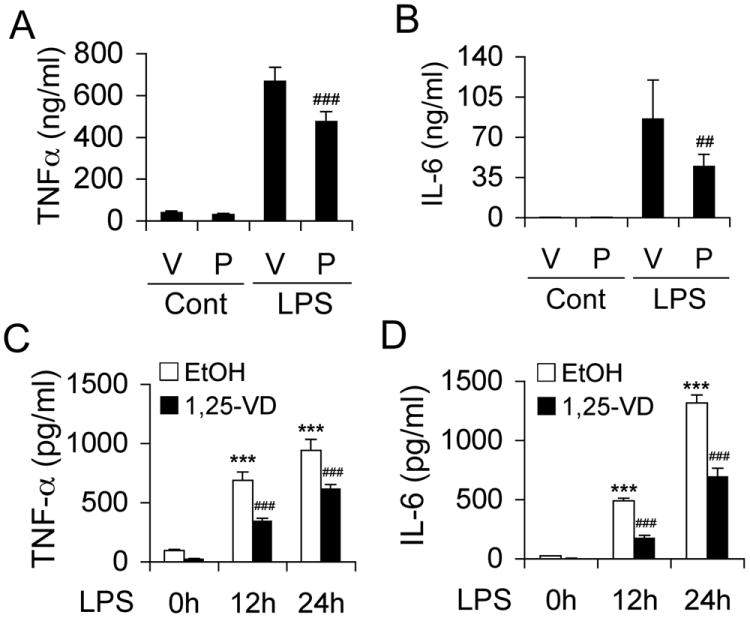
Figure 2.

VDR activation inhibits LPS-induced inflammatory reaction. (A and B) WT mice were pretreated with vehicle (V) or paricalcitol (P, 200 ng/kg, daily i.p.) for one week before being challenged with saline control or LPS (20 mg/kg). Serum TNFα (A) or IL-6 (B) concentration was measured at 0 and 24 hours after LPS injection. ## P<0.01; ### P<0.001 vs. V; n=7. (C and D) WT BMDMs were pretreated with ethanol (EtOH) or 1,25(OH)2D3 (1,25-VD, 20 nM) for 24 hours before being exposed to LPS (100 ng/ml). The time course of TNFα (C) and IL-6 (D) release into the media was determined by ELISA. ***, P<0.001 vs. 0 hour; ### P<0.001 vs. corresponding EtOH; n=3.
