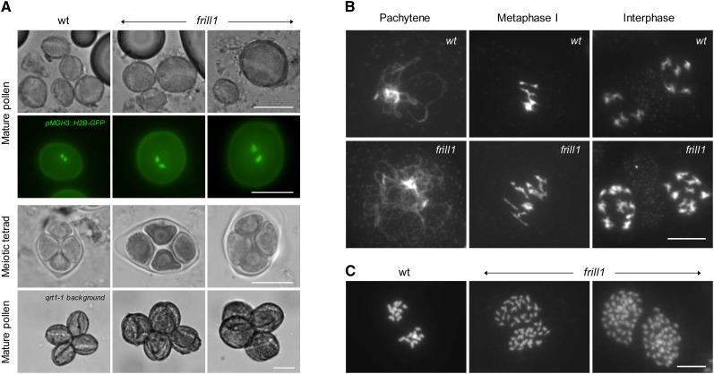
Figure 6.
Cytological Analysis of frill1 Male Sporogenesis.
(A) Bright-field microscopy of mature pollen isolated from wild-type (wt) and frill1 flowers at anthesis and fluorescent visualization of sperm cells in mature pollen grains using the ProMGH3-H2B-GFP construct. Analysis of the male meiotic products in wild-type and et2 anthers through cytological tetrad stage meiocyte examination and mature pollen grain observations in the qrt1-1−/− mutant background. Bars = 20 µm.
(B) DAPI-stained meiotic chromosome spreads of wild-type and frill1 male sporogenesis at different stages of the meiotic cell cycle. Bar = 10 µm.
(C) Chromosome spreading of somatic cells in premeiotic frill1 flower buds shows the ectopic formation of endomitotic polyploid cells. Bar = 5 µm.
[See online article for color version of this figure.]

