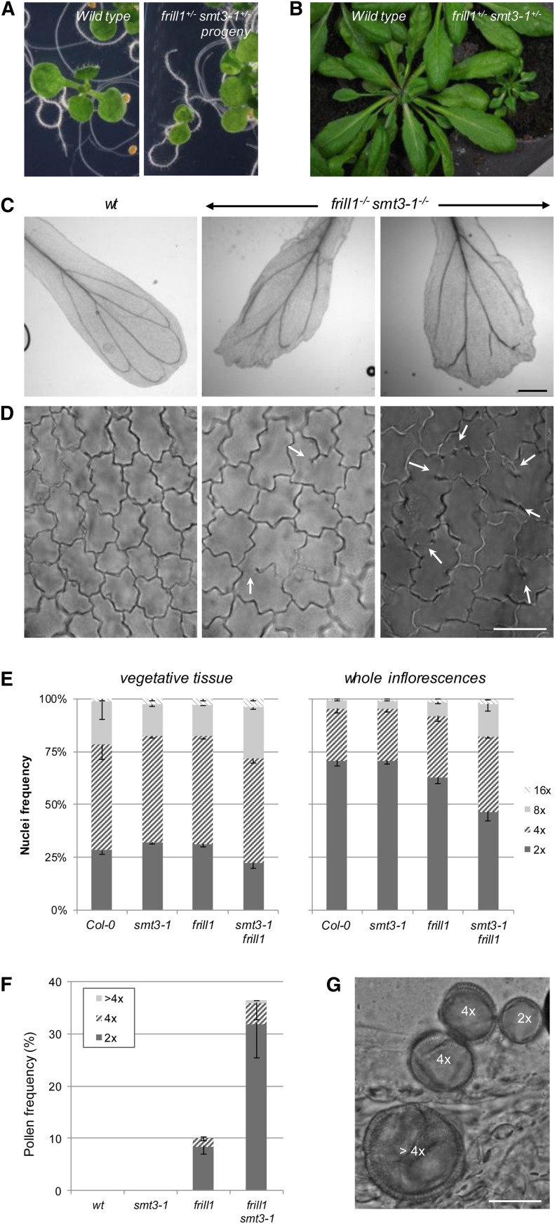
Figure 9.
Phenotypic Characterization of Double frill1−/− smt3-1−/− Mutants.
(A) and (B) General morphology of wild-type and frill1−/− smt3-1−/− 7-d-old seedlings and 4-week-old plants.
(C) Cytological analysis of petal vasculature and distal margin morphology in the wild type (wt) and frill1−/− smt3-1−/−. Bar = 250 µm.
(D) Epidermal cell wall morphology in wild-type and frill1−/− smt3-1−/− petals. Arrows indicate cell wall stubs and incomplete cell walls. Bar = 20 µm.
(E) Quantitative analysis of the endoploidy pattern of vegetative leaf tissue and whole inflorescences of 6-week-old plants. For each sample, three biological replicates were analyzed and error bars represent sd.
(F) Quantitative analysis of larger pollen grain formation in flowers of 6-week-old plants. For each sample, three biological replicates were analyzed and error bars represent sd.
(G) Normal sized diploid (2x), enlarged tetraploid (4x), and jumbo polyploid (>4x) pollen grains in frill1−/− smt3-1−/− flowers. Bar = 20 µm.
[See online article for color version of this figure.]

