Figure 2.
TIE1 Is a Novel Nuclear Transcription Repressor.
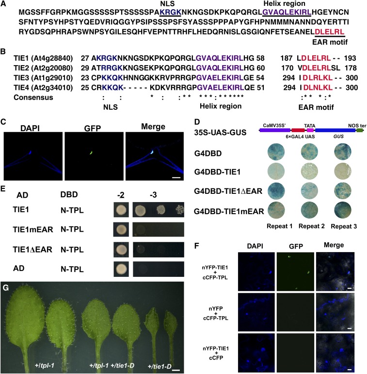
(A) The amino acid sequence of TIE1 (At4g28840). TIE1 contains a monopartite nuclear localization signal (NLS) (underlined, blue), a helix region (underlined, purple), and an EAR motif (underlined, red).
(B) Partial amino acid sequence alignment of TIE1, TIE2 (At2g20080), TIE3 (At1g29010), and TIE4 (At2g34010). They all contain a nuclear localization signal, a helix region, and an EAR motif. Asterisks indicate identical residues and colons indicate similar residues.
(C) TIE1 is localized in the nucleus. Trichome cell from a leaf of 35S-TIE1-GFP-3 transgenic line expressing TIE1-GFP fusion protein. From left to right, the staining of nucleus by DAPI, fluorescence of GFP, and merge of DAPI and GFP. Bar = 25 µm.
(D) TIE1 is a transcription repressor. The transcription activity of TIE1 was tested in tobacco leaves using a GAL4/UAS-based system. CaMV 35S', 35S promoter without TATA box; 6×GAL4 UAS, six copies of GAL4 binding site (UAS); NOS ter, terminator of nopaline synthase gene; G4DBD, GAL4 DNA binding domain; G4DBD-TIE1, G4DBD fused with TIE1; G4DBD-TIE1ΔEAR, G4DBD fused with TIE1ΔEAR in which the EAR motif was deleted; G4DBD-TIE1mEAR, G4DBD fused with TIE1mEAR in which the three conserved Leu residues of the EAR motif was mutated into Ser residues.
(E) TIE1 interacted with TPL protein through the EAR motif in yeast two-hybrid assays. AD, activation domain; DBD, DNA binding domain; TIE1mEAR, mutated TIE1 in which the three conserved Leu residues of the EAR motif was mutated into Ser residues. TIE1ΔEAR, deleted TIE1 in which the EAR motif was deleted; N-TPL, the TPL N terminus including residues from 1 to 188. Transformed yeasts were spotted on control medium (-2) or selective medium (-3) in 10-, 100-, and 1000-fold dilutions. The empty vectors were used as controls.
(F) BiFC analysis of the interaction between TIE1 and TPL. From left to right, DAPI staining showing the nuclei, GFP fluorescence, and merge of DAPI and GFP. Bars = 20 µm.
(G) Genetic interaction between tpl-1 and tie1-D. From left to right, the 4th and 5th leaves from 20-d-old +/tpl-1, +/tpl-1 +/tie1-D double mutant, and +/tie1-D. Bar = 1 mm.

