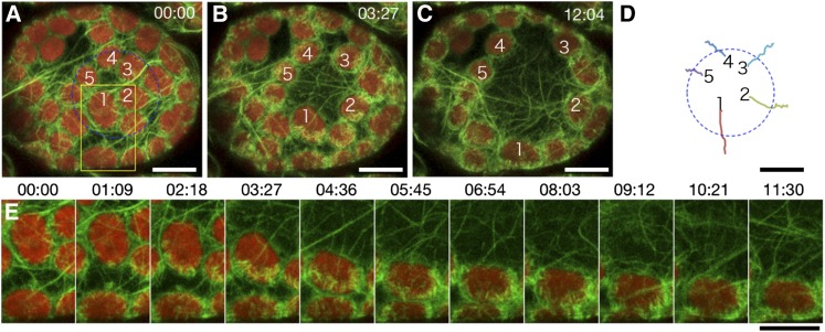
Figure 3.
Reconstruction of Cp-Actin Filaments on the Leading Edge of Chloroplasts during Avoidance.
(A) to (C) Serial images of chloroplasts during avoidance induced with 458-nm laser scans inside the blue circle (20 µm in diameter) during the intervals between the image acquisitions. Time-lapse images were collected at 34.5-s intervals for 12 min and 4 s. The images are false-colored to indicate GFP (green) and chlorophyll (red) fluorescence. The cp-actin filaments on the chloroplasts before (A), during (B), and after (C) the avoidance response are shown. The time points (minutes:seconds) at image acquisition are shown in the top right corner of the pictures. See also Supplemental Movie 5 online for the full time-lapse series. Bars = 10 µm.
(D) The paths of the individual chloroplasts that moved. The centers of the representative chloroplasts, 1 to 5 in (A) to (C), were traced. Bar = 10 µm.
(E) Enlarged and more precise time-lapse images of chloroplast 1 in the yellow inset of (A). Note that the reconstruction of cp-actin filaments always occurred in the front side of the moving chloroplast, even if the chloroplast turned during the movement. Bar = 10 µm.

