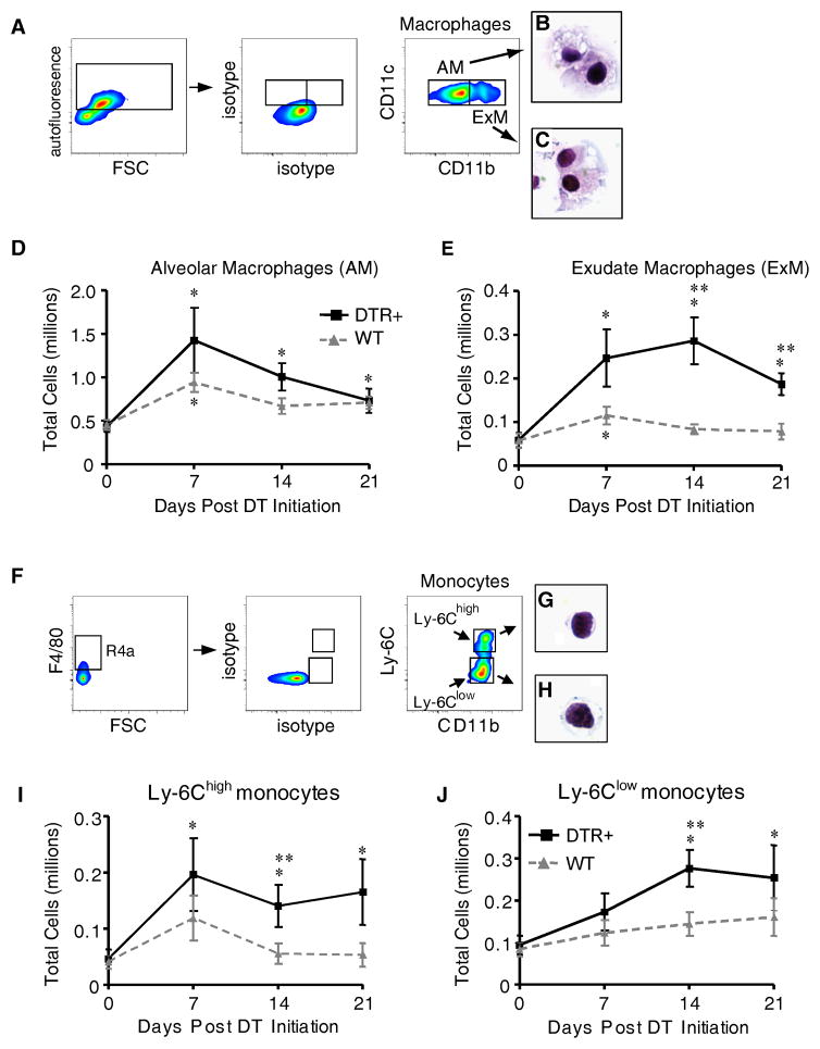
Figure 2. Exudate macrophages and Ly-6Chigh monocytes accumulate in mice with targeted injury to type II AEC.
Diphtheria toxin was administered per protocol for 14 days to either WT (DTR−) or DTR+ mice. (A–J) Lung cells isolated from untreated mice (D0) and at 7, 14, and 21 days after the onset of DT treatment were stained with specific antibodies and analyzed by flow cytometric analysis as described in Materials and Methods and Supplemental Figure S1. (A) Gating strategy used to identify AM and ExM among lung leukocytes obtained from DTR+ mice after 14 days of DT-treatment. Initial gating (not shown) on CD45+ lung leukocytes excludes debris, lymphocytes, and CD11c-negative cells (refer to Figure S1). Within the CD11c-postive populations, a plot of FL-3 vs. FSC (left dot plots) distinguishes larger autofluorescent macrophages (AF+) from smaller non-autofluorescent cells. Amongst AF+ macrophages, plots of isotype controls (middle dot plots) and CD11c vs. CD11b (right dot plots) distinguish AM (as AF+ CD11c+ CD11b−, gate AM) and ExM (as AF+ CD11c+ CD11b+, gate ExM). (B, C) Representative photomicrographs of AM (B) and ExM (C) obtained by flow sorting of lung leukocytes at D14 (100x objective; H&E stain). (D, E) Total numbers of AM (D) and ExM (E) present in the lungs of WT or DTR+ mice at each timepoint. (F) Gating strategy used to identify Ly-6Clow and Ly-6Chigh monocytes among lung leukocytes obtained from DTR+ mice after 14 days of DT-treatment. Initial gating (not shown) on CD45+ lung leukocytes excludes debris, lymphocytes, granulocytes, and CD11c-positive cells (refer to Figure S1). Within the SSClow CD11c-negative population (left dot pot), a gate of F4/80 vs FSC identifies F4/80-positive monocytes. Next, plots of isotype controls (middle dot plot) and Ly-6C vs. CD11b (right dot plot) identify Ly-6Chigh monocytes (gate Ly-6Chigh) and Ly-6Clow monocytes (gate Ly-6Clow). (G, H) Representative photomicrographs of Ly-6Chigh monocytes (G) and Ly-6Clow monocytes (H) obtained by flow sorting of lung leukocytes at D14 (100x objective; H&E stain). (I, J) Total numbers of Ly-6Chigh monocytes (I) and Ly-6Clow monocytes (J) present in the lungs of WT or DTR+ mice at each timepoint. Total macrophage and monocyte numbers were calculated by multiplying the frequency of each population (using the gating strategy described above) by the total number of CD45+ lung leukocytes at each time point. Data represent mean ± SEM of 8–12 mice assayed individually per time point. * p<0.05 ANOVA vs. Day 0 (untreated) of mice of the same DTR expression profile; ** p<0.05 when values from WT mice or DTR+ mice were compared against each other at the designated time point.

