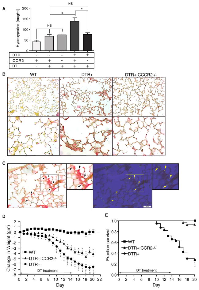
Figure 4. CCR2 expression mediates lung collagen deposition, weight loss, and increases mortality following injury to type II AEC.
(A) Diphtheria toxin was administered for 14 days per protocol to: 1) WT mice (DTR−:CCR2+/+), 2) CCR2-deficient mice (DTR−:CCR2−/−), 3) DTR+ mice (DTR+:CCR2+/+), and 4) DTR+:CCR2−/−mice. An additional group of control WT mice received daily intraperitoneal injections of PBS for 14 days. Lungs were harvested on day 21 and analyzed for hydroxyproline content. Results are reported as the mean concentration in μg/ml ± SEM (n = 6 to 8). P values <0.05 between groups were considered significant. (B) Photomicrographs taken of lung sections obtained from DT-treated WT mice (left panels), DTR+ mice (middle panels), and DTR+:CCR2−/− mice (right panels) at Day 21 (one week after completing DT treatment) and stained with picrosirius red to identify collagen (top panels, 20x objective, bottom panels 40x objective). Note the increase in subepithelial collagen staining (red color) observed in lungs from DTR+ mice which is not observed in WT mice and minimal in DTR:CCR2−/− mice. (C) Photomicrographs (20x objective) of picrosirius-stained lung sections obtained from DT-treated DTR+ mice (D21) viewed by bright field (left panel and inset) and cross-polarized (right panel and inset) microscopy. Black arrows (left panels) identify regions of increased picrosirius staining. Yellow arrows (right panels) identify corresponding regions displaying birefringence (yellow fluorescence) indicative of co-aligned collagen. (D, E) Weight loss (D) and survival (E) in WT, DTR+, and DTR+:CCR2−/− mice treated with DT per protocol for 14 days. * p<0.05 vs DT-treated WT mice; ‡ p<0.05 vs DT-treated DTR+:CCR2−/− mice.

