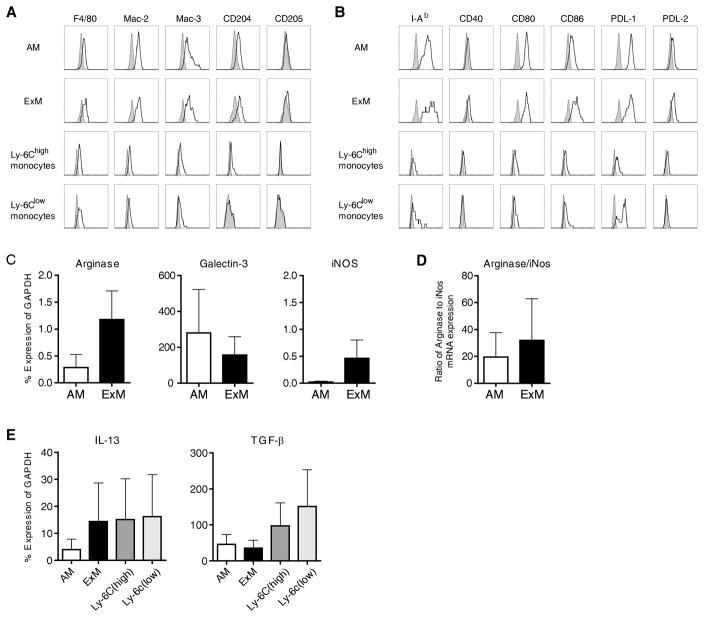
Figure 5. Immunophenotype of ExM and Ly-6Chigh monocytes in mice with type II AEC injury.
(A, B) Representative histograms (four-decade log scale) of ExM and Ly-6Chigh monocytes pooled from four individual DTR+ mice at D14 of DT-treatment displaying their expression of (A) macrophage-associated proteins (F4/80, Mac-2, Mac-3, CD204, CD205) and (B) MHC Class II (I–Ad) and costimulatory molecules (CD40, CD80, CD86, PDL-1, and PDL-2). Shaded histogram, isotype staining; open histogram, specific staining. The experiment was repeated once with similar results. (C–E) Designated subsets of lung macrophages and monocytes were purified (≈95% by cell sorting) from pooled populations of lung leukocytes obtained from DTR+ mice (n=5 mice) at D14 of DT-treatment. mRNA obtained from each subset was assessed by qRT-PCR. Specific gene expression is expressed as a percentage of GAPDH (Methods). Values represent the average ± SEM from three independently-performed experiments. (C, D) AM and ExM expression of genes associated with (C) alternative (Arginase-1, Galectin-3) or classical (iNOS) macrophage activation. (D) Ratio of Arginase:iNOS gene expression (as calculated for each experiment). (E) Macrophage and monocyte expression of profibrotic cytokine genes, IL-13 and TGF-β.

