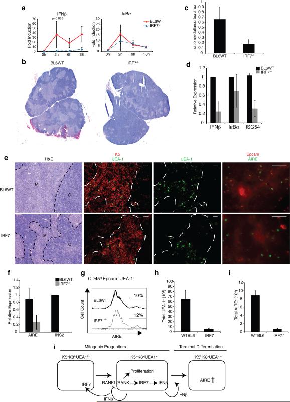
Figure 5. IRF7 is necessary for thymic IFNβ expression and mTEC development.
a) Thymic stromal cells from WT or IRF7-/- mice were purified and stimulated with 500 ng/mL RANKL for the indicated time points. Expression of IFNβ and IκBα mRNAs was determined by qPCR. b) Thymic architecture in WT and IRF7-/- mice revealed by H&E staining of thymic sections (bar = 500 μm) c) Ratio of medullary to cortical cellularity in thymic sections from WT and IRF7-/- mice (data collected using ImageJ (NIH) and represent avg +/-sd of 3 sections each from 3 mice (p<0.05). d) mRNA levels of IFNβ, Iκbα, and ISG54 in thymic stromal cells from WT or IRF7-/- mice as determined by qPCR. e) Thymic architecture in WT and IRF7-/- mice as revealed by H&E staining of thymic sections (left panels), Keratin 5 and UEA-1 expression (middle panels, bar = 100 μm), UEA-1 only, as well as EpCAM and AIRE expression (right panels, bar = 50 μm) f) Relative mRNA levels of AIRE and INS2 in purified stromal cells from WT and IRF7-/-thymi measured by qPCR (n = 3). g) Histogram shows percentage of AIRE+ cells in the CD45loEpcam+UEA-1hi gate of thymic stromal cells from WT and IRF7-/- mice. h) Total number of UEA-1hi cells as determined by flow cytometric analysis of CD45loEpCAM+UEA-1hi mTEC populations from WT and IRF7-/- thymi. i) Total number of AIRE+ cells based on flow cytometric analysis of CD45loEpCAM+UEA-1hiAIRE+ mTEC populations in WT and IRF7-/- thymi (n = 4). j) Hypothetical model of IFNβ production and function in the thymus.

