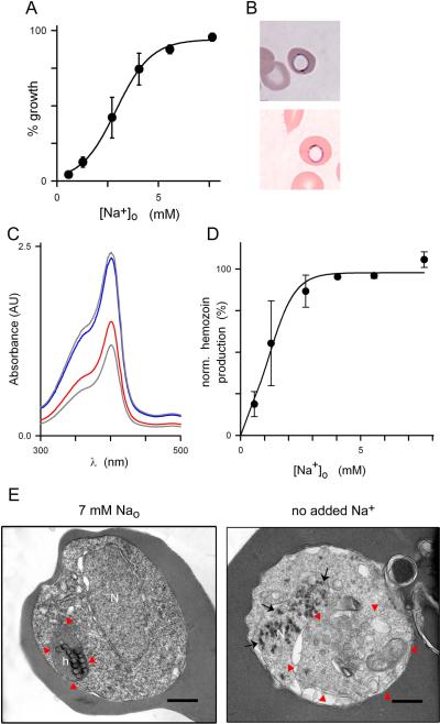
Fig. 3.
Low Na+ concentrations are required. (A) Normalized parasite growth over 72 h in 4suc:6KCl with 10% dialyzed serum and indicated Na+ concentrations, revealing a sigmoidal dose response for Na+ requirement. Mean ± S.E.M. of 9 replicates from 3 experiments. (B) Photomicrograph of Giemsa-stained trophozoite-infected cells after 24 h cultivation in 4suc:6KCl with 10% dialyzed serum without Na+ supplementation. Two parasites are shown with a rim of blue stain and central clearing. (C) Absorbance scan showing reduced β-hematin production in trophozoites cultivated in Na+-deficient medium (red trace); supplementation with 7 mM Na+ restores β-hematin production (blue trace), yielding levels similar to those observed with standard RPMI-based medium (upper gray trace). Positive control, 20 μM chloroquine (lower gray trace). AU, arbitrary units. (D) Mean ± S.E.M. β-hematin production vs. external Na+ concentration, normalized to zero for control culture with 20 μM chloroquine. (E) Transmission electron micrographs showing trophozoite-infected erythrocytes cultivated in 4suc:6KCl with 10% dialyzed serum with or without Na+ supplementation to a final 7 mM concentration (left and right panels, respectively). Red arrowheads demarcate the parasite digestive vacuole; black arrows show electron dense spots in nucleus when Na+ is not added. N, nucleus; h, normal hemozoin crystals; scale bars, 0.5 μm. Images are representative of 89 cells from two separate experiments.

