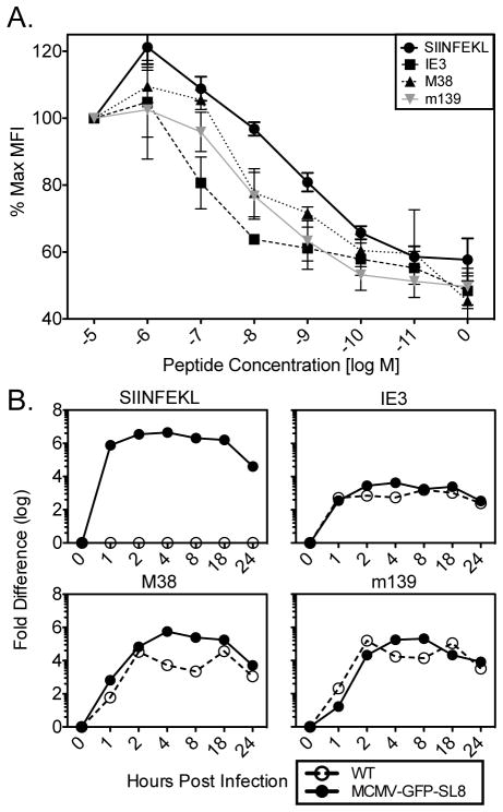
Figure 5. MHC binding affinity of MCMV epitopes and kinetics of expression.
(A) RMA-S cells were incubated with the indicated concentrations of peptide for 2hrs at 25°C and an additional 2hrs at 37°C, then washed and stained for H2-Kb expression. Experiment was done twice. Shown is the normalized mean fluorescence intensity of class I MHC on the surface of cells. (B) Murine embryonic fibroblasts were infected with the indicated viruses and RNA was harvested at the time points listed on the y-axis. cDNA was made in parallel with no reverse-transcriptase controls for each sample, and qRT-PCR was done for the indicated gene products. No signal was obtained from the no reverse transcriptase controls. Experiment was done twice.

