FIGURE 2.
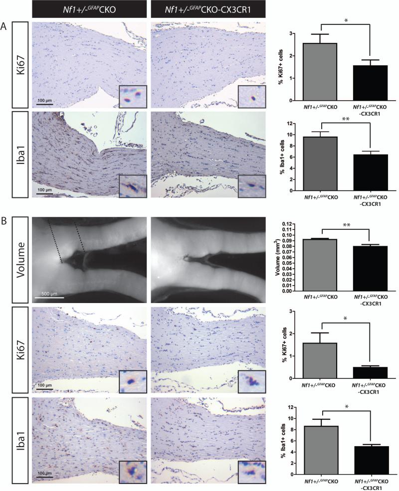
Targeted reduction of CX3CR1 expression reduces microglia content and optic glioma proliferation. (A) Optic nerves from Nf1+/-GFAPCKO-CX3CR1 mice (n=9) exhibit a 39% reduction in the percent of Ki67+ cells relative to Nf1+/-GFAPCKO (n=9) at 6 weeks of age (p = 0.0463) as well as a 33% reduction in the percent of Iba1+ cells (p = 0.0086). (B) 3-month-old Nf1+/-GFAPCKO-CX3CR1 mice (n=10) have smaller optic nerve volumes relative to Nf1+/-GFAPCKO mice (n=10, 13% reduction, p = 0.0039) and exhibit a 65% reduction in proliferation (%Ki67+ cells; p = 0.0358) as well as a 42% reduction in the percentage of Iba1+ microglia (p = 0.0114), relative to Nf1+/-GFAPCKO mice. Dashed lines delineate the representative area of the optic nerves used to calculate volume. Insets show representative positively-labeled cells. *, p<0.05; **, p<0.01.

