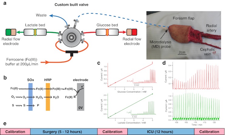
Fig. 1.

The microdialysis probe is inserted into the tissue flap subcutaneously and sutured into place, shown in the photograph in (a) before the flap has been detached from the blood vessels. The dialysate is introduced to the rsMD injection valve via low-volume FEP connection tubing (blue dotted line). The reaction is mediated by ferrocene monocarboxylate, which is perfused at 200 μL/min using an HPLC pump. The enzyme is loaded onto membranes placed before a glassy carbon electrode which is held at 0 V (b). The current is recorded as peaks, where amplitude is related to concentration as shown in the calibration curve in (c). Raw data of a successful arterial anastomosis (indicated by the dotted grey line) are shown in (d). A time line indicating the calibration protocol throughout monitoring is shown in (e)
