
Figure 4.
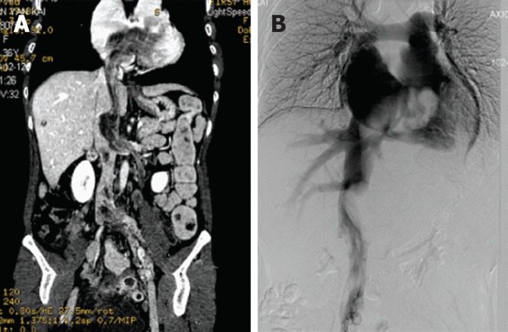
Coronal plane computed tomography and digital subtraction angiography image of intravenous leiomyomatosis. A: Preoperative coronal plane computed tomography demonstrates a mass was present in the left ovarian vein, left renal vein, left external and common iliac vein, as well as within the inferior vena cava, extending into the right atrium; B: Digital subtraction angiography image demonstrates an irregular mass filling defect from the inferior vena cava to the right atrium.