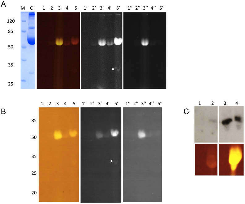
Figure 1. Flu-PAGE and Flu-Blot analyses of human serum.
Normal human serum (lanes 1, 1′ and 1″) and serum samples labelled with fluorescein (lanes 2, 2′ and 2″), fluorescein-boronic acid (lanes 3, 3′ and 3″), rhodamine (lanes 4, 4′ and 4″) and rhodamine-boronic acid (lanes 5, 5′ and 5″) in a 12% non-denaturing polyacrylamide gel (A) and Eastern blot (B). The gels and blots were imaged with Dark Reader® (lanes 1–5), UV (365 nm, with orange filter (595 nm); lanes 1′–5′) and UV (365 nm, with green filter (537 nm); lanes 1″–5″). The Coomassie stained control lane is shown in the left panel, labelled as C. The asterisks indicate an extra band present in the rhodamine-boronic acid labelled sample (lane 5′) on both the Flu-PAGE and Flu-Blot. (C) shows the Western blot analysis of glucose incubated HSA after 0 (lanes 1 and 2) and 28 days (lanes 3 and 4) using anti-AGE antibodies. Lanes 1 and 3 correspond to unlabelled samples, whereas lanes 2 and 4 are labelled with fluorescein-boronic acid.

