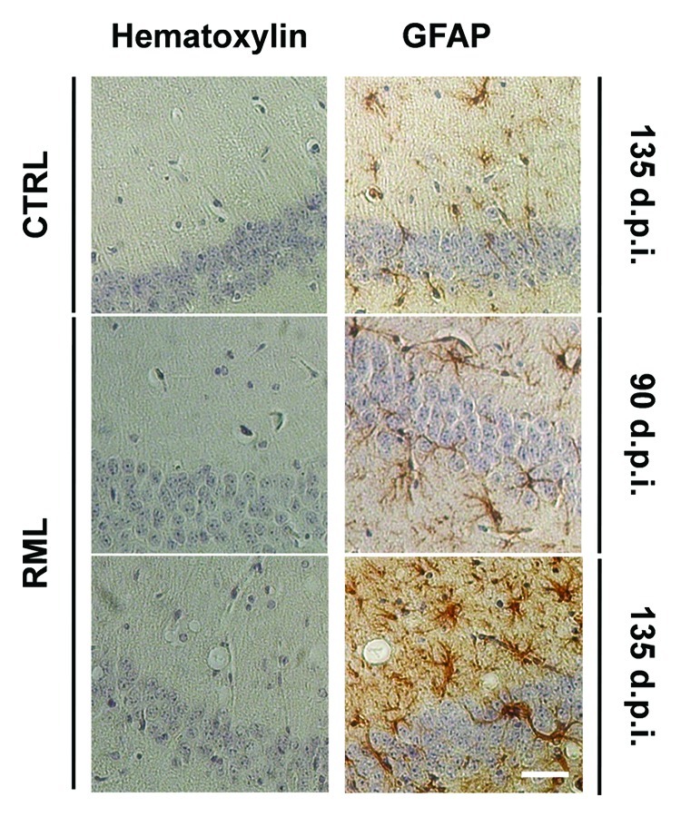
Figure 2. Progressive astrocytosis and spongiosis in the hippocampus of scrapie-infected mice. Coronal sections from saline-treated (CTRL) and RML-infected animals (i.c.) at 90 and 135 d.p.i were stained with hematoxylin alone or hematoxylin with the astrocytic marker Glial Fibrillary Acidic Protein (GFAP). Stainings highlight spongiosis and pronounced astrocytosis in the hippocampus of RML-infected mice at 135 d.p.i. Images are representative of 3 animals. Scale bar, 50μm.
