
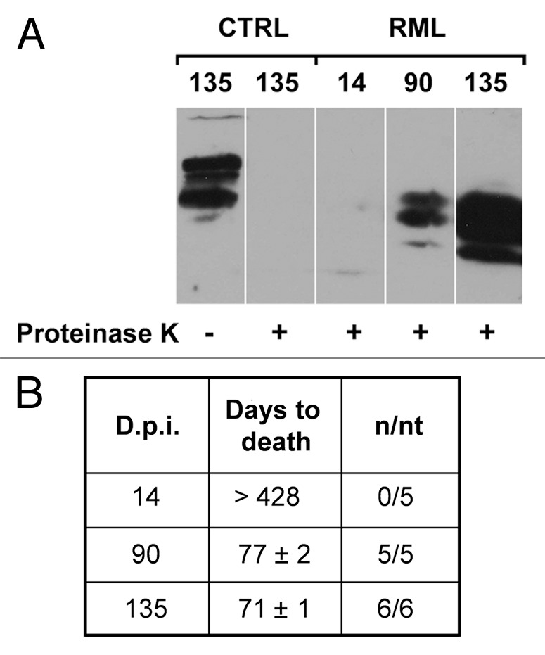
Figure 3. PrPSc accumulation and prion titers in the hippocampus of RML-infected mice. (A) Hippocampal homogenates from saline-treated (CTRL) or RML-infected mice (i.c.) at 14, 90 and 135 d.p.i. were treated in the presence (+) or in the absence (-) of proteinase K to distinguish native PrPC from the proteinase K-resistant PrPSc and analyzed by western blotting (n = 3). (B) Prion titers in the hippocampus of infected animals at 14, 90 and 135 d.p.i. were determined by inoculating homogenates i.c. into CD-1 indicator mice. Data highlight a progressive increase in the amount of PrPSc and infectivity in the hippocampi of RML-infected mice at 135 d.p.i. n/nt = Number of mice with scrapie/ total number of mice inoculated.