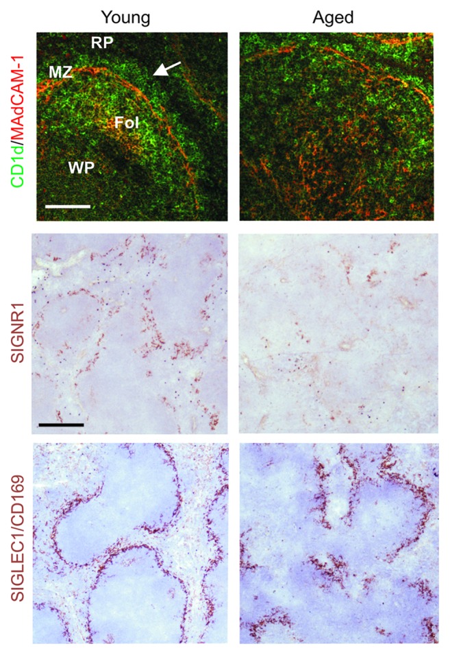
Figure 4. The microarchitecture of the splenic MZ is disrupted in the spleens of aged mice. Left-hand-panels; In the spleens of young mice MADCAM1-expressing sinus-lining cells form a distinct barrier between the MZ and the white pulp (WP, arrow). RP, red pulp; Fol, FDC-containing B cell follicle. Within the MZ abundant CD1d-expressing MZ B cells (upper panels, green) continually shuttle between MZ and B cell follicles as they transfer immune complexes to FDC. Two distinct populations of macrophages also reside in the MZ: SIGNR1-expressing MZ macrophages, and SIGLEC1/CD169-expressing MZ metallophilic macrophages (middle and lower panels, brown). In the spleens of aged mice the distribution and density of these cells are all severely disrupted when compared with young mice (right-hand panels). Upper panels, scale bar = 50 µm; middle and lower panels, scale bar = 100 µm. Adapted from Brown et al.34
