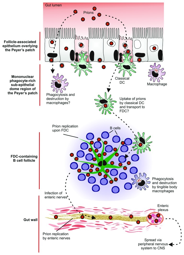
Figure 5. A potential cellular relay that mediates prion neuroinvasion after oral exposure. After ingestion of a contaminated meal, prions appear to be actively transcytosed into Peyer’s patches by M cells and enterocytes in the follicle-associated epithelium. In the absence of M cells neuroinvasion is blocked suggesting that M cells are the important sites of prion uptake from the gut lumen. The prions are subsequently acquired by mononuclear phagocytes (macrophages and classical DC) in the sub-epithelial dome of the Peyer’s patches. Current hypotheses suggest classical DC, in contrast to macrophages, act as ‘Trojan horses’ and carry the prions to the FDC in the B cell follicles. The prions then infect and replicate upon FDC. Following their expansion upon FDC, prions subsequently infect enteric nerves. The prions then spread to the CNS via the peripheral nervous system (both sympathetic and parasympathetic).

An official website of the United States government
Here's how you know
Official websites use .gov
A
.gov website belongs to an official
government organization in the United States.
Secure .gov websites use HTTPS
A lock (
) or https:// means you've safely
connected to the .gov website. Share sensitive
information only on official, secure websites.
