
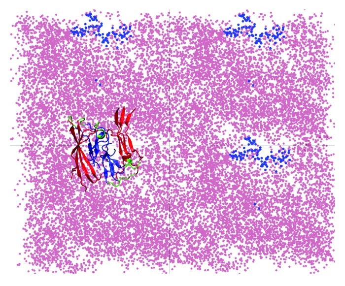
Figure 2. Repositioning of lipid headgroups due to interactions with the Aβ40 protofilament segment facilitates the access of the hydrophobic C-terminal β-strands into the lipid tail region (top view; see also Fig. 1). Here, four top-view periodic images of the simulation box are shown for a typical trajectory. The dots represent the positions sampled by the center of mass (COM) of the lipid P atoms (pink) and the COM of the C-termini (blue) during the 45 to 150 ns trajectory segment when the C-termini are associated with the lipid head groups. A representative Aβ9–40 octamer structure is shown in only one periodic image.