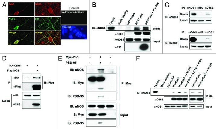
Figure 1. Cdk5 Interacts with NOS1. (A) Co-localization of NOS1 and Cdk5 in rat cerebrocortical neurons. NOS1 and Cdk5 were detected by immunocytochemistry in cultures after 14 d in vitro. Cdk5 and NOS1 staining superimpose to show co-localization in deconvolved images. Rat cerebrocortical neurons were also stained with anti-p35 antibody. No staining was observed when primary antibodies were omitted. (B) GST-pull down assay. GST-fusion protein or GST alone (GST-empty) was incubated with rat brain homogenates for 3–5 h and then pulled down with GSH beads. The precipitates were subjected to immunoblot and probed with NOS1 antibody. GST-Cdk5 or GST-Cdk5 plus His-p35, but not GST protein alone, pulled down NOS1 (lanes 4 and 5). (C) Co-immunoprecipitation (Co-IP) of NOS1 and Cdk5 from rat brain homogenates. Homogenates were immunoprecipitated with Cdk5 or NOS1 antibody and then probed with these antibodies. Anti-HA antibody was used as a control. (D) Interaction of Cdk5 and NOS1 is independent of p35. HEK293T cells were transfected as indicated, lysed, and immunoprecipitated with anti-Flag or anti-HA antibody. Precipitates and total lysates were subjected to immunoblotting. (E) p35 does not directly interact with NOS1. HEK293-NOS1 cells were transfected as indicated, lysed and, immunoprecipitated with anti-Myc antibody. Precipitates and total lysate were subjected to immunoblotting. (F) Co-IP of NOS1 and Cdk5 in HEK293-NOS1 cells. HEK293-NOS1 cells transfected with HA-tagged Cdk5, HA-tagged mtCdk5 (non-nitrosylatable mutant), or pcDNA3.0 were treated with 5 μM A23187 in the presence or absence of 1 mM N-nitro-l-arginine (NNA), immunoprecipitated with HA antibody, and then probed with NOS1 antibody.

An official website of the United States government
Here's how you know
Official websites use .gov
A
.gov website belongs to an official
government organization in the United States.
Secure .gov websites use HTTPS
A lock (
) or https:// means you've safely
connected to the .gov website. Share sensitive
information only on official, secure websites.
