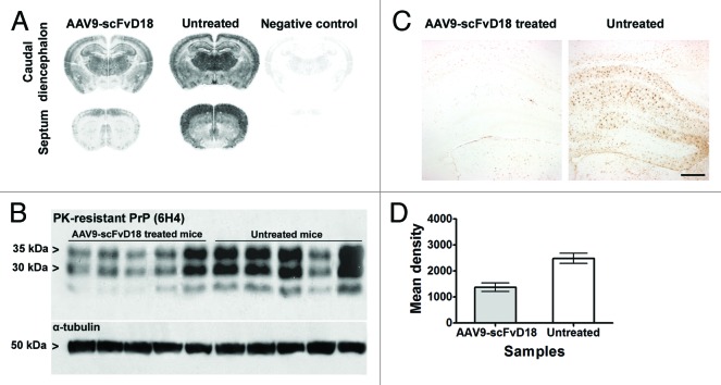
Figure 4. PK-resistant PrPSc deposition in scrapie infected mice at an early stage of disease (166 d.p.i). Histoblots with anti-PrP antibody (6H4) showed lower PK-resistant PrPSc deposition in the brain of AAV9-scFvD18 treated mice than that of untreated animals (A). Western blot analysis (B) with anti-PrP antibody (6H4) of PK-digested brain homogenates from AAV9-scFvD18 treated and untreated mice and the relevant densitometric analysis (D) demonstrate statistically significant lower levels of PK-resistant PrPSc in the brain of AAV9-scFvD18 treated mice (p = 0.005; double tailed, unpaired, t-test). Astrocytic activation (C) parallel the PK-resistant PrPSc deposition, and is much more severe in untrated animals. Scale bar = 20 μm (all microphotographs are at the same magnification).

An official website of the United States government
Here's how you know
Official websites use .gov
A
.gov website belongs to an official
government organization in the United States.
Secure .gov websites use HTTPS
A lock (
) or https:// means you've safely
connected to the .gov website. Share sensitive
information only on official, secure websites.
