
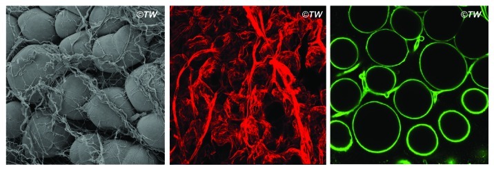
Figure 1. Peri-adipocyte collagens. Left: the scanning electron micrograph of mouse inguinal fat pads; the group of round adipocytes are surrounded with collagen bundles. Middle: immunofluorescent staining of type I collagen (red); thick bundles of type I collagen surrounding the group of adipocytes as well as thinner fibers of type I collagen enwrapping individual adipocyte is displayed. Right: immunofluorescent staining of type IV collagen (green); type IV collagen is found as a component of basement membrane that enwraps each adipocytes; type IV collagen can also be found as the basement membrane underneath the layer of vascular endothelial cells.