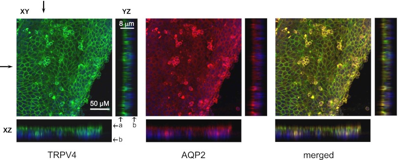
Figure 10.
Systemic GSK1016790A treatment normalizes subcellular distribution of TRPV4 and AQP2 in freshly isolated CD-derived cyst monolayers. Representative confocal plane micrographs (axes are shown) and corresponding cross-sections (arrows) of three-dimensional stacks of TRPV4 localization (pseudocolor green), AQP2 localization (pseudocolor red), and the combined image for a CD-derived cyst monolayer from PCK453 rat treated with GSK1016790A for 1 month. Nuclear staining with DAPI is shown by pseudocolor blue. a, apical side; b, basolateral side; DAPI, 4',6-diamidino-2-phenylindole.

