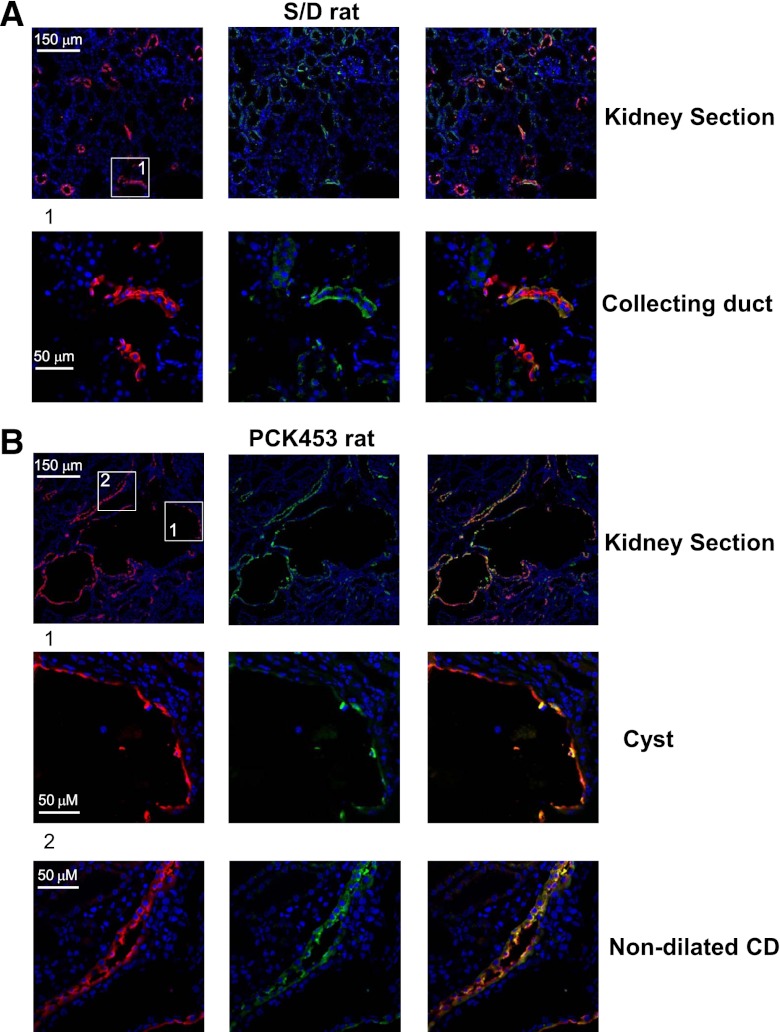
Figure 5.
Subcellular TRPV4 localization is different in cysts and nondilated CDs in PKC453 rats. (A) Immunofluorescence of a kidney section from Sprague-Dawley (S/D) rat. A representative low-magnification 10-μm-thick kidney section (top row) shows staining patterns for TRPV4 (pseudocolor green), AQP2 (pseudocolor red), and the combined image. The area defined by a rectangle is shown below with a higher magnification. (B) Immunofluorescence of kidney section from a PCK453 rat. A representative low-magnification 10-μm-thick kidney section (top row) shows staining patterns for TRPV4 (pseudocolor green), AQP2 (pseudocolor red), and the combined image. Areas defined by rectangles and containing a cyst (middle) and a nondilated CD (bottom) are shown below with a higher magnification.

