
Figure 9.
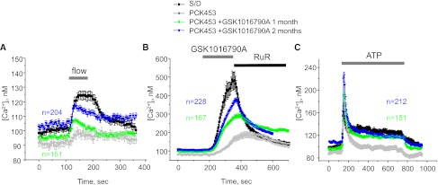
Mechanosensitive [Ca2+]i signaling is restored in CD-derived cyst cells of GSK1016790A-treated PCK453 rats. The average time course of [Ca2+]i changes in response to an abrupt 10× elevation in flow (A), application of GSK1016790A (30 nM) followed by 2 μM Ruthenium red (B), and application of 10 μM ATP (C) for individual cells within freshly isolated CD-derived cyst monolayers from PCK453 rats treated with GSK1016790A for 1 (green) and 2 months (blue), respectively. The time courses of control CD cells from Sprague-Dawley (S/D) rats (black) and CD-derived cyst monolayers from untreated PCK453 rats (gray) are reproduced from Figure 2A. Numbers of all individual recordings for each condition are shown in the respective color. Durations of treatments are shown on the top with bars.