Figure 2.
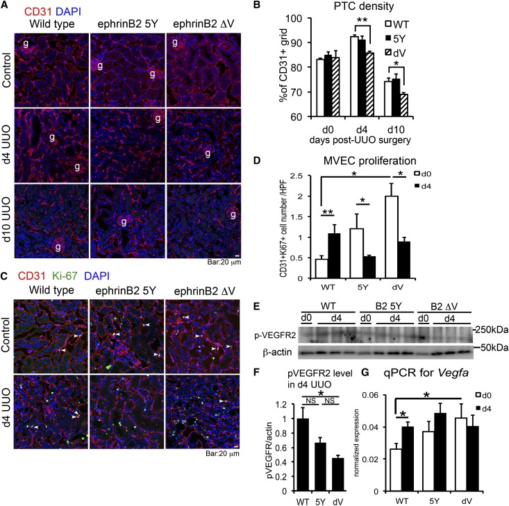
Defective PDZ-dependent ephrinB2 signaling promotes progressive capillary rarefaction and impairs angiogenesis after UUO kidney injury. (A) Immunofluorescence images of CD31+ MVECs in control, d4, and d10 UUO kidneys of WT, ephrinB2 5Y (5Y), and ephrinB2 ΔV (dV) mice. (B) Quantification of PTC density. n=3–4 per group per time point. (C) Immunofluorescence images of CD31+ Ki-67+ proliferative MVECs in control and d4 UUO kidneys of WT, ephrinB2 5Y, and ephrinB2 ΔV mice. Arrowheads indicate double-positive cells. (D) Quantification of CD31+ Ki-67+ proliferative MVECs. n=3 per group per time point. (E) Western blot for p-VEGFR2, β-actin in d0 (normal) and d4 UUO kidneys of WT, ephrinB2 5Y, and ephrinB2 ΔV mice. (F) The protein band intensity of p-VEGFR2 in d4 UUO kidneys assessed by densitometry. Intensity of p-VEGFR2 band is normalized by β-actin band intensity. Note that p-VEGFR2 expression is reduced in ephrinB2 ΔV compared with WT. n=3 per group. (G) qPCR showing VEGFA transcriptional level in d0 and d4 UUO kidneys of WT, ephrinB2 5Y, and ephrinB2 ΔV mice. n=3 per group. *P<0.05; **P<0.01. g, glomerulus; NS, no significant difference. Scale bar, 20 μm.

